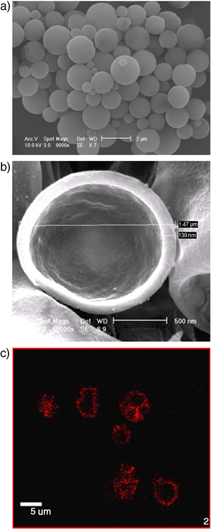
Figure 3.
Images of drug loaded agents. a) SEM after fabrication (Mag. = 9000x, Size bar = 2 µm). b) SEM after sample ruptured by sonication (Mag. = 50000X, Size bar = 500 nm). c) Fluorescent confocal micrograph showing Dox within the agent’s shell (Mag=100X, Size bar= 5 µ, (only larger UCA are visible using fluorescent microscopy)). Agent shown is a PLA agent with 3% (g Dox/g PLA) loaded within the shell of the agent. Morphology, core, and shell thicknesses were consistent with all three loading methods and all drug payloads.

