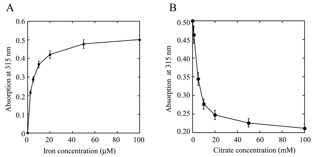
Figure 2. Iron binding activity of hIscA1 in vitro.
A) Iron binding titration of hIscA1. The iron-depleted hIscA1 (50 µM) was incubated with Fe(NH)2(SO4)2 (0 to 100 µM) in the presence of dithiothreitol (2 mM) at 37°C for 30 min, followed by re-purification of hIscA1. The absorption amplitude at 315 nm of re-purified hIscA1 was plotted as a function of the Fe(NH)2(SO4)2 concentration in the incubation solution. B) Iron binding competition of hIscA1. The iron-saturated hIscA1 (100 µM) was incubated with sodium citrate (0 to 100 mM) in the presence of dithiothreitol (2 mM) at 37°C for 30 min, followed by re-purification of hIscA1. The absorption amplitude at 315 nm of re-purified hIscA1 was plotted as a function of the sodium citrate concentration in the incubation solution. The results are the means ± SD from three independent experiments

