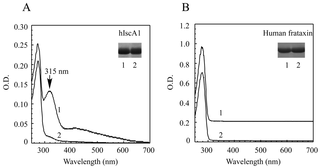
Figure 3. Iron binding activity of hIscA1 in E. coli cells.
A) The UV-visible absorption spectra of hIscA1 purified from the E. coli cells grown in the M9 minimal medium supplemented with (spectrum 1) or without (spectrum 2) 50 µM ferrous ammonium sulfate. The protein concentration was approx. 50 µM. The insert is a photograph of the SDS/PAGE gel of hIscA1 purified from the E. coli cells grown in the M9 minimal medium supplemented with (lane 1) or without (lane 2) 50 µM ferrous ammonium sulfate. B) The UV-visible absorption spectra of human frataxin purified from the E. coli cells grown in the M9 minimal medium supplemented with (spectrum 1) or without (spectrum 2) 50 µM ferrous ammonium sulfate. The protein concentration was approx. 20 µM. The insert is a photograph of the SDS/PAGE gel of frataxin purified from the E. coli cells grown in the M9 minimal medium supplemented with (lane 1) or without (lane 2) 50 µM ferrous ammonium sulfate. The results are representatives from three independent experiments.

