Figure 5.
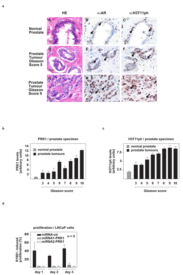
PRK1 and H3T11ph levels positively correlate with the malignancy of prostate cancer and control tumour cell proliferation. (a), Immunohistochemical staining of AR and H3T11ph in human normal and tumour prostate. AR (B, E, H) and H3T11ph (C, F, I) immunoreactivity is detected in the secretory epithelium of normal prostate (B, C, arrows) and prostate carcinoma cells (E, F, H, I, arrows). Hematoxilin-eosin (HE) stained sections are shown in A, D, and G. All sections were taken from the same radical prostatectomy specimen (magnification: ×250). (b, c) The correlation of elevated PRK1 and H3T11ph levels with high Gleason score in a panel of 111 human prostate carcinomas is highly significant: r=0.499, p<0.001 (b) and r=0.450, p<0.001 (c). Normal prostate specimens (n=20) are included as a control. (d) In LNCaP cells, miRNA-mediated PRK1 knockdown severely reduces R1881-induced cell proliferation. Bars represent mean +SD (n≥4).

