
Figure 2. Suppression of EGFR expression promotes apoptosis in H. pylori-infected gastric epithelial cells.
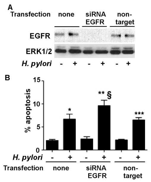
ImSt tansfected with EGFR siRNA or non-targeting siRNA were infected with H. pylori for 24 h. A: EGFR expression levels were analyzed by Western blot analysis of cellular lysates using an anti-EGFR antibody. An anti-ERK1/2 antibody was used as a loading control. B. Apoptosis was detected using TUNEL assay as described in Figure 1. Apoptosis was determined by counting at least 500 cells. The percentage of cells undergoing apoptosis is shown. *, **, and ***p < 0.001 vs uninfected cells in each cell line, § p < 0.001 vs non-transfected cells infected with H. pylori.