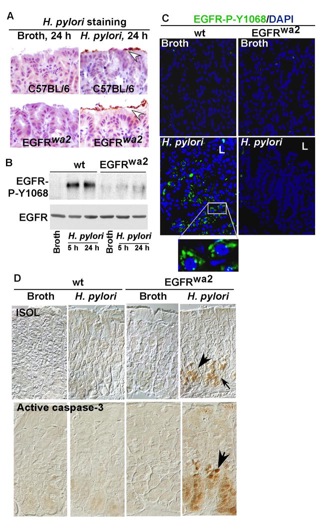
Figure 3. Loss of EGFR kinase activity promotes gastric epithelial cell apoptosis in mice infected with H. pylori.
Wt and EGFR wa2 (EGFR kinase defective) mice on a C57BL/6 background were infected with H. pylori (109 cfu/mouse) for 5 or 24 h. Paraffin-embedded stomach tissues (A, C–D) and gastric mucosal lysates (B) were prepared. A. Immunostaining with anti-H. pylori antiserum was performed to detect the presence of H. pylori (arrowheads). B: EGFR Y1068 phosphorylation was detected by Western blot analysis of gastric mucosal lysates as described in Figure 1. C: EGFR Y1068 phosphorylation was detected using immunostaining with anti-EGFR Tyr1068 and FITC-labeled secondary antibody. Green represents positive labeling (L: Luminal side). D: Apoptosis was detected by ISOL staining and active caspase-3 immunostaining using an anti-active caspase-3 antibody. Apoptotic nuclei were labeled with peroxidase and developed with DAB and observed using DIC microscopy. Brown nuclei (arrows) represent ISOL or activated caspase-3 positive staining cells. n = 5–7 mice for each condition.

