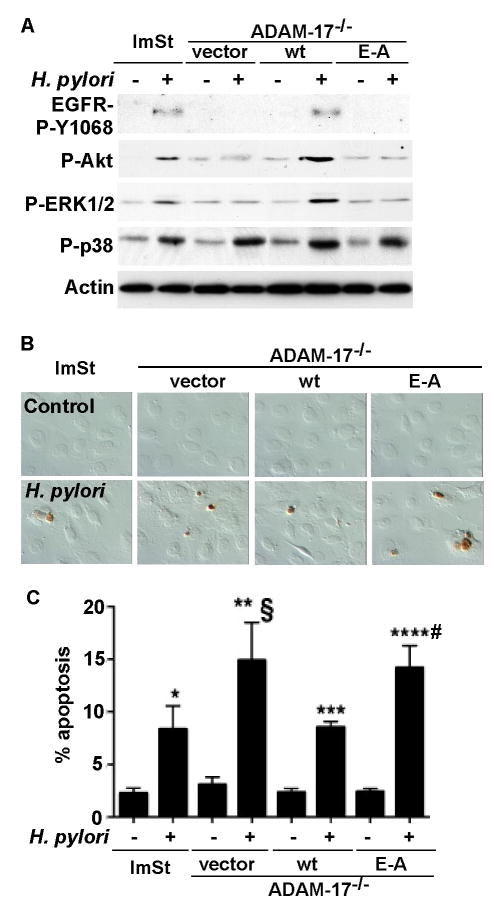
Figure 4. ADAM-17 mediates H. pylori-induced activation of EGFR and is required for an anti-apoptotic effects.
Cells were infected with H. pylori for 90 min (A) or 24 h (B and C). EGFR Y1068 phosphorylation was analyzed by Western blot analysis of cellular lysates (as described in Figure 1), activation of Akt using anti-phospho (P)-Ser 473 Akt, ERK1/2 MAPK activation using anti-phospho (P)-Thr183/Tyr185 ERK1/2, and p38 using anti-phospho (P)-Tyr180/182 p38 antibodies. B–C: Cells were fixed for TUNEL assay and apoptotic nuclei were labeled with peroxidase and developed with DAB and observed using DIC microscopy. Brown staining represents apoptotic nuclei (B). Apoptotic cells were enumerated by counting at least 500 cells. The percentage of cells undergoing apoptosis is shown (C). *, **, *** and ****p < 0.001 vs uninfected cells in each cell line. § and # p < 0.001 vs ImSt and wt ADAM-17 transfected cells infected with H. pylori.

