Figure 5.
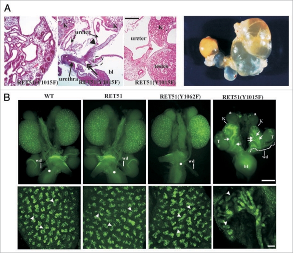
Complex CAKUT phenotype RET51(Y1015F) due to supernumerary ureters and decreased branching. (A) Histological and gross features of RET-Y1015F-PLCγ signaling mutant. The H&E-stained pictures at P0 show cystic hypodysplastic kidney (left), dilated ureters and possible distal stricture (middle) and failure of testes to separate from the urinary system (right). The gross photograph shows massive megaureter/hydroureter and barely any recognizable kidney parenchyma in 4 week old RET(Y1015F) mutant mice. (B) Whole mount Dolichos Biflorus Agglutinin-FITC (DBA-FITC) staining of E15.5 kidneys. Top, Normal honeycomb pattern of ureteric bud branching, single ureters entering the bladder (white dot), and separation of Wolffian duct (wd) from the ureter was evident in kidneys (K) of mice expressing WT, RET51 and RET51(Y1062F). In RET51(Y1015F) animals, note bilateral small kidneys with supernumerary UBs (arrows) that enter the mesenchyme but show decreased branching and failed to separate from the wd (white dashed line). The testes (T) were also abnormally positioned. Bottom, Imaging of the above kidneys by confocal microscopy clearly depicts the dramatic deficit in branching morphogenesis (arrowheads indicate branch points) in homozygous RET51(Y1015F) kidneys. Scale bar: 100 µm. Adapted and modified from Jain et al.42 with permission.

