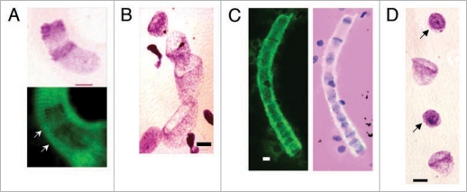
Figure 2.
Bell shaped, metakaryotic nuclei in syncytia. (A) Feulgen purple stained stacking bell-to-bell symmetrical amitotic fission of bell shaped nuclei (upper) imposed over green Feulgen fluorescent image (lower) showing sarcomeric striations (arrowed) of syncytial walls (human fetal spinal cord, 9 wks). (B) Feulgen purple stained bell shaped nuclei in syncytium illustrating variety of nuclear dimensions (human fetal gut, 7 wks). (C) Green fluorescent image of a single syncytium with bell shaped nuclei (left image) and the same image merged with the image of Feulgen purple stained nuclei showing positions of the nuclei in syncytium (human fetal gut, 7 wks). Note, both “kissing cup” and “stacked cup” amitotic figures and bell shaped nucleus apparently emerging from end of syncytium. (D) Section of a Feulgen purple stained syncytium showing a portion of a larger series of eight condensed spherical nuclei (arrowed) between bell shaped nuclei interpreted as the result of synchronous asymmetrical amitoses (human fetal gut, 7 wks). Scale bar, 5 µm.

