Figure 3.
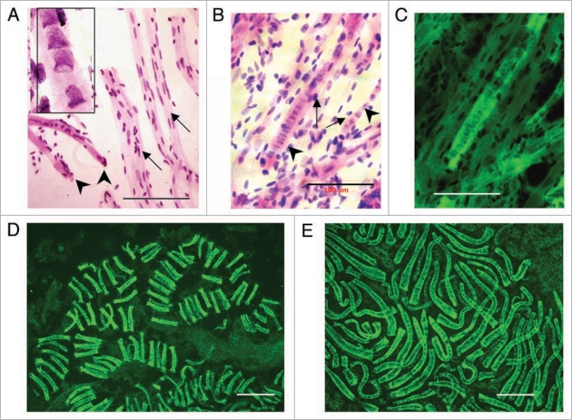
Syncytial clusters in different human fetal meta-organs. (A) Cardiac unstriated muscle at 10 weeks: Feulgen purple stained tubular syncytia (left low corner, arrows) with bell shaped nuclei in central position (zoomed image, left upper corner, 100x). Tubular syncytia are striated with bell shaped nuclei and thus distinct from differentiated cardiac unstriated muscle fibers (arrows) with closed elliptical nuclei aligned with surface of muscle fiber. (B) Skeletal striated muscle of thigh at 10 weeks: Feulgen purple stained tubular syncytia (arrows) lying in parallel with striated muscle fibers (arrowed) with sarcomeric structures with closed elliptical nuclei apparently externally associated to muscle fibers. (C) Same image as (B) merged with green Feulgen fluorescent image. Fluorescence from metakaryotic tubular syncytia is more intense than from differentiated muscle fibers. (D) Spinal cord ganglia at 9 weeks. Multiple clusters of tubular syncytia with 16 bell shaped nuclei each, green Feulgen fluorescence superimposed on purple stained Feulgen image. (E) Brain at 9 weeks. Clusters of syncytia with ∼16–32 bell shaped nuclei each stained as in (C and D). Scale bar, 100 µm.

