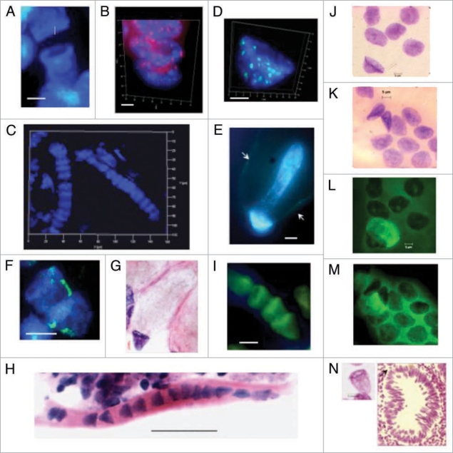
Figure 6.
Bell shaped nuclei observed with various techniques in tissues, tumor and cell culture preparations. (A) DAPI stained DNA (blue) syncytial bell shaped nuclei in human fetal spinal cord ganglia, 8–9 wks. (B) DAPI staining (blue) and pan-telomeric FISH labeling (Cy3, red) of bell shaped nuclei in mouse fetal spinal cord ganglia tissue, 16.5 days. (C) DAPI stained syncytia with multiple bell shaped nuclei as seen in 3D imaging [Z-stack, ‘Apotome’], human fetal spinal cord ganglia, 9 wks. (D) High resolution 3D image of DAPI stained bell shaped nucleus and pan-centromeric FISH staining (FITC green), human colon adenocarcinoma, 68 yrs. (E) DAPI stained bell shaped nucleus dividing asymmetrically (bell to cigar shaped nucleus) in human colon adenocarcinoma, 68 yrs. [Arrows indicate the walls of the balloon shaped cytoplasm through which new eukaryotic nuclei migrate]. (F) DAPI stained bell shaped nucleus dividing symmetrically with segregation of pair of chromosomes 18 stained by FISH (FITC green), fetal spinal cord ganglia, 8–9 wks. (G) H&E staining of metakaryotic extra syncytial cell, colon adenocarcinoma, 68 yrs. (H) H&E staining of syncytial bell shaped nuclei, spinal cord, 8–9 wks. (I) Acridine orange stained syncytial bell shaped nuclei, spinal cord, 8–9 wks. (J and L) Feulgen stained DNA (purple) bell shaped nuclei appearing in human adenocarcinoma cell line. (K and M) Feulgen fluorescent (green) images of (J and L), respectively showing unidentified fluorescent material in cytoplasm of all cells of bell shape and with a small fraction of cells with spherical nuclei. (N) Feulgen stained bell shaped nucleus as seen in 30 microns snap frozen tissue section of human colon polyp, 27 yrs. Arrow indicates position of the nucleus relativel to aberrant crypt. Bar scales, 5 µm (A–N) except 50 µm in (H).

