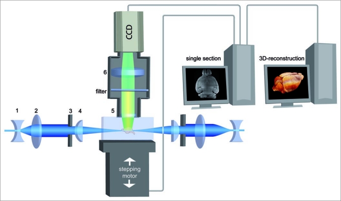
Figure 1.
Principle of UM. A transparent specimen is illuminated perpendicular to the observation pathway by a laser forming a thin sheet of light. Concave lens (1), convex lens (2), slit aperture (3), cylindrical lens (4). Thus, fluorescent light is only emitted by a thin plane and no out of focus light has to be excluded by a pinhole like in confocal microscopy. The emitted fluorescence light is projected to a camera target by an objective, while the excitation light is blocked by a matched optical filter. Objective (5), tube lens (6). By moving the specimen chamber through the light sheet a stack of images is obtained. Afterwards, a 3D-reconstruction is calculated by software.

