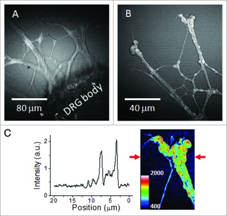
Figure 2.
CA RS images of unstained live neurites growing from the DRG body cultured in 1% CS gel matrix. CA RS images of (A) live neurites at the proximity to the DRG body and (B) neurites and bulbs observed around the end of a neuronal growth cone. (C) Signal profile along a line indicated by the arrows in the inset image.

