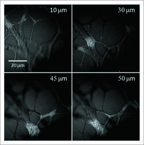
Figure 3.
3-D structure of unstained neurites growing in a 1% CS gel matrix inspected by a laser-scanning CA RS microscope. Representative images at different depths show the 3-D distribution of the neurite growth. The number marked in each image indicates the depth relative to the bottom layer of neurite observed in the field of view.

