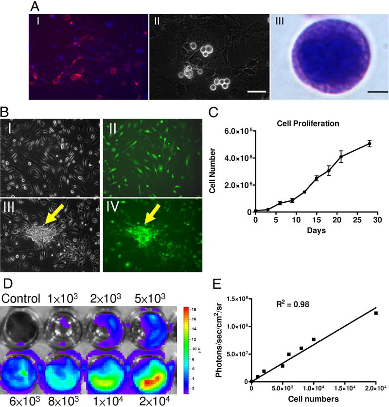
Figure 1. Isolation and culturing of cardiac stem cells.
(A) Ventricular cells from L2G85 transgenic mice stained for Sca-1 (red) (I). After a period ranging from 1 to 3 weeks, phase-bright cells migrated over a layer of fibroblast-like cells (II). The phase-bright cells were collected and Giemsa stain showed the cell with large nucleus (III). Scale bar=50μm (i, ii), 2μm (III). (B) Subculture and in vitro differentiation of phase-bright cells. Morphology of CSCs (I) cultured in poly-D-lysine-coated plates and expressing GFP (II). With cardiac differentiation medium, some CSCs can form cardiac sphere (arrow) with GFP expression (III, IV). (C) Proliferation curves show linear growth of cultured CSCs over a one month period. (D) & (E) Ex vivo imaging analysis of CSCs show increasing bioluminescence signals with cell numbers (r2=0.98)

