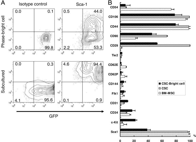
Figure 2. Characterization of surface markers in cardiac stem cells.
Quantification by FACS analysis of CSC phase-bright cells and its derived cells. Bone marrow mononuclear cells derived mesenchymal stem cells (BM-MSC) were used as control. (A) CSCs express robust GFP both on phase-bright CSCs and their subcultures. After 2–3 passages, there was upregulation of Sca-1. Lower panels showed Sca-1 antibody isotype control. (B) Quantitative analysis of cell markers expression by FACS. CSCs express high level mesenchymal stem cells markers, CD29, CD90, CD44, and CD106. After subculturing, Sca-1 up-regulated but c-Kit, CD34, CD31, and CD144 markers all down-regulated. Compare to BM-MSC, CSCs express less CD54, but higher CD29 and CD90. All FACS experiments were performed in triplicates.

