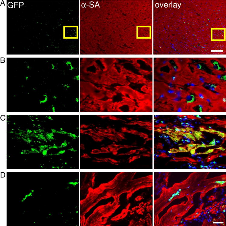
Figure 7. Tracking of grafted CSCs by immunofluorescence.
(A – B) CSCs within the recipient myocardium 3 days after injection shown at low and high magnification. (C–D) Transplanted CSCs can differentiate and integrate with host myocardium as confirmed by GFP and α-sarcomeric actin (α-SA) double staining. At day 14, CSCs could differentiate into cardiomyocytes as confirmed by α-SA and GFP double stainings (Figure 7C). However, this population became significantly decreased when the tissues were examined at day 28 (Figure 7D), which is also consistent with the decrease in bioluminescence signals over this period of time. Scale bar=100μm (A), 20μm (B, C, D).

