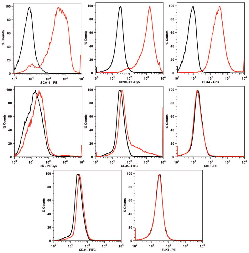
Fig. 1.
Adipose-derived stromal cells from passages 3 to 5 expressed identical cell surface antigens when analyzed by fluorescence-activated cell sorting. Adipose-derived stromal cells were positive for the cell surface markers Sca-1, CD90, and CD44 and negative for all lineage markers (CD4, CD8, CD11b, B220, GR-1, and TER-119), CD45, c-kit/CD117, CD31, and Flk-1. These data are congruent with the marker profile of murine-derived stromal cells described in previous studies. Data are expressed as a histogram plot, with black representing isotype control and red representing experimental.

