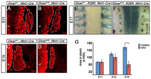
Fig. 4. Dicer is required for sensory nervous system survival but not neuronal differentiation.
(A–D) Neuronal differentiation in the DRG was examined by immunofluorescent analysis of expression of the pan-neuronal marker Tuj1 at E11 and E15 of development. At E11, the DRG of (A) control and (B) Dicerfx/fx; Wnt1-Cre embryos have formed and express Tuj1 (A). At E15, the DRG of control embryos (C) continues to grow while DRG of Dicerfx/fx; Wnt1-Cre embryos (D) fail to expand. (E–F) The result of Dicer loss on DRG survival and patterning late in development was examined in E17 embryos by tracing NC derived cells using β-galactosidase expression from the R26R locus. A comparison of a dorsal view of the DRG from control (E) and Dicer mutant (F) embryos shows that the DRG are maintained in mutant embryos but the ganglia size of mutant embryos is reduced and project axons. (G) To determine when during development the DRG fails to expand in mutant embryos, the cross sectional area of ganglia was calculated using the pan-neural marker Tuj1 to mark neurons. At E11 and E13, there is no significant difference between the control and mutant ganglia (P=0.967 and P=0.209 respectively). By E15, there is a significant decrease in the size of the ganglia in mutant embryos (P=0.003).

