Figure 1.
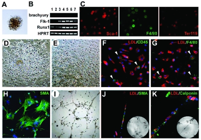
Molecular characteristics, hematopoietic and vascular potential of BL-CFC derived from E10.5 mouse AGM region. (A) Morphology of a typical blast colony. (B) Expression of Brachyury, Flk-1 and Runx1 in seven representative blast colonies determined by nested RT-PCR. (C) Immunofluorescence staining of Sca-1, F4/80 and Ter119 in blast colonies. Morphology of individually plucked blast colonies in the liquid expansion system after 48 h (D) and 10 days (E) of incubation. (F–G) DiI-Ac-LDL incorporation (red) combined with CD45 or F4/80 staining (green) of the adherent cells. Arrowheads, CD45 positive in H or F4/80 positive in I. (H) α-SMA+ staining (green) of the adherent cells. (I) Tube-like structures in Matrigel of the adherent cells. (J–K) DiI-Ac-LDL incorporation (red) combined with α-SMA or Calponin staining (green) of the tube-like structures. Inserts show the corresponding bright fields. Original magnification: ×40 (I), ×100 (D, E), and ×200 (A, C, F–H, J, K).

