
Figure 5.
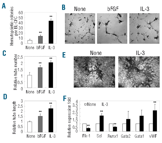
Increased hematopoietic and vascular potential of IL-3-treated BL-CFC from the E10.5 AGM region. (A) IL-3- and bFGF-stimulated BL-CFC gave rise to increased numbers of hematopoietic CFC. (B–D) A Matrigel-based test revealed that IL-3- and bFGF-stimulated BL-CFC have enhanced tube-forming capacity both with regards to the number and length of the tubes. (E) CD31 staining of blast colonies co-cultured with OP9 for 10 days. Increased tube formation was detected when IL-3 was added during blast colony formation. (F) Real-time PCR analysis of BL-CFC cultures to examine IL-3-mediated molecular changes. Original magnification: ×40 (B, E). The results represent means ± s.e.m. Significance was determined using the Student’s t- test: **, P<0.01, compared with the control data.