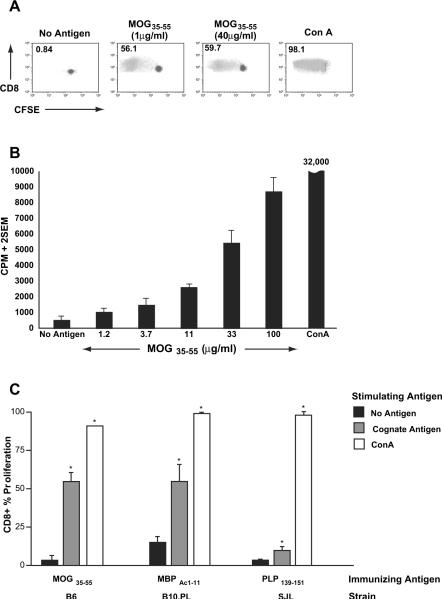
Fig. 2.
Purified CD8+ T cells confirm neuroantigen-specific reactivity. Untouched CD8+ T cells were purified from bulk splenocytes of MOG-immunized B6 mice and subjected to either CFSE-based proliferation assays (A) or 3H-thymidine-based proliferation assays (B). In each case, purified CD8+ T cells were incubated with irradiated, CD8-depleted splenocytes from OVA323–339-immunized mice. The numbers in panel A represent % proliferation and demonstrate robust MOG35–55-specific CD8+ T cell responses. Panel B demonstrates dose-response to MOG35–55. Results are representative of over 10 independent experiments. Panel C shows CFSE-based proliferation assays conducted on splenocytes from B6, B10.PL and SJL mice immunized with MOG35–55/CFA (B6), MBPAc1–11/CFA (B10.PL) or PLP139–151/CFA (SJL). The bars represent % proliferation of purified CD8+ T cells and are representative of two to three independent experiments. Proliferation responses to cognate antigens and Con A were significantly greater than background (*p < 0.01).

