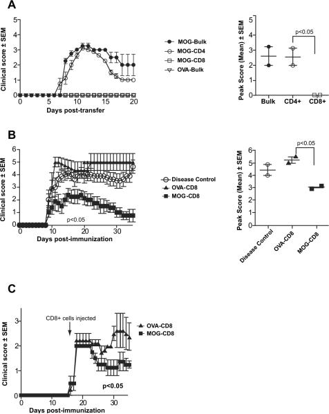
Fig. 3.
MOG35–55-specific CD8+ T cells show a regulatory, but not pathogenic, role in wildtype B6 EAE. (A) Splenocytes from MOG35–55/CFA-immunized female B6 mice were harvested at day 25 post-immunization and stimulated in vitro for 72 h with MOG35–55 and mIL-2. Following incubation, either bulk splenocytes (MOG-Bulk) or magnetically purified CD4+ (MOG-CD4+) or CD8+ (MOG-CD8+) cells were injected intravenously into naïve wildtype B6 mice. As controls, splenocytes were harvested from OVA323–339/CFA-immunized mice (OVA-Bulk) and similarly adoptively transferred following in vitro activation. Adoptive EAE was monitored and is depicted as mean clinical score ± SEM (left panel). Analyses of mean peak scores showed no difference between MOG-CD4+ and MOG-Bulk cells, although MOG-specific-CD8 cells were consistently incapable of transferring disease (right panel). These results are representative of over 8 independent experiments. (B) Splenocytes from MOG35–55-or OVA323–339-immunized mice were harvested at day 25 post-immunization and were cultured in vitro with their respective antigens. CD8+ T cells were purified by magnetic sorting and injected intravenously into naïve wildtype B6 mice (“day 1”). One day later (“day 0”), EAE was induced with MOG35–55/CFA. EAE scores for the various groups are shown on the left. Analyses of mean peak scores showed significant differences between the mean peak scores of OVA-CD8 and MOG-CD8 groups. These results are representative of five independent experiments. (C) EAE was induced in B6 mice with MOG35–55/CFA. Mice were split into two groups at day 13 of disease (randomized based on disease scores) and received either in vitro activated OVA323–339- or MOG35–55-stimulated CD8+ T cells. Mice were then monitored for disease (observation period of 40 days) in a blinded manner. These results are representative of three independent experiments.

