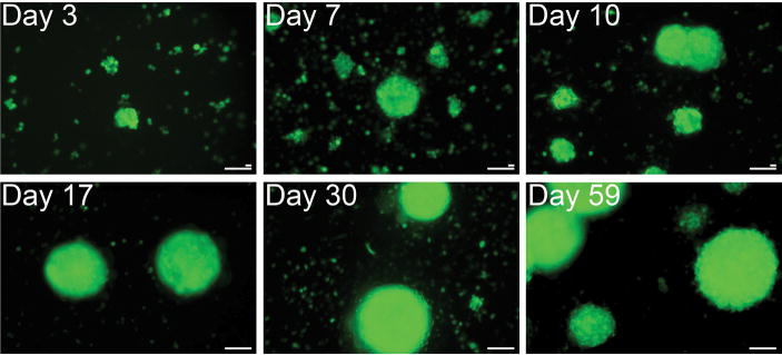
Figure 1. Formation and growth of myospheres over time.
Immediately after isolation myosphere cultures were transduced with a lentiviral vector expressing GFP, the formation of myospheres were monitored at various time points after isolation: 3, 7, 10, 17, 30, and at 59 days. The size of the myospheres formed ranged from 50 μm to over 300 μm. Larger scale bar represents 100 μm, and the smaller bar, 20 μm.

