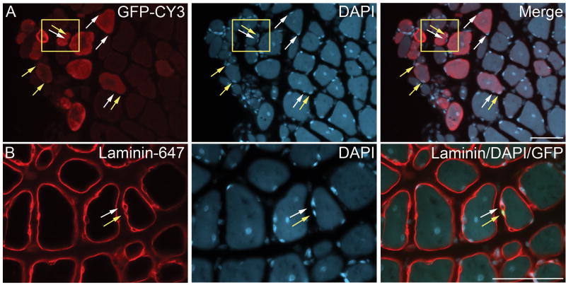
Figure 7. Myospheres contribute to mononuclear cells in regenerating muscle.
Donor myosphere cells expressing GFP were injected into the TA muscle of CTX injured mdx mice. Two weeks after injection the mice were sacrificed. Cross-sections of the injected TA muscles are shown. (A) GFP staining detected by CY3 shown in red (left panel), DAPI staining (nuclear, center panel), and the merger of GFP and DAPI (right panel). Yellow arrows indicate GFP positive nuclei from the donor cells and white arrows indicate GFP negative nuclei of the recipient mouse. (B) Laminin staining detected by Alexa 647 shown in red (left panel), DAPI staining (nuclear, center panel), and a merger of laminin and DAPI staining with GFP (right panel). The yellow arrow shows a donor mononuclear cell found within the basal lamina (GFP positive, shown in green) and the white arrow shows a mononuclear cell from the recipient mouse (GFP negative). Scale bars represents 50 μm.

