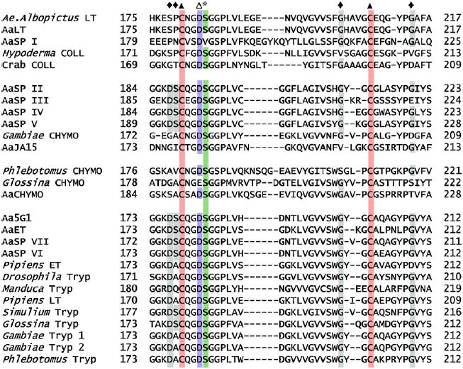
Fig. 3.

Protein alignment of serine proteases. Protein alignment of all serine proteases used in this analysis from positions ~170–220. Numbering is based on bovine α-chymotrypsinogen. Residues of importance are represented as follows; (*) Ser195 catalytic triad residue, (◆) accessory catalytic residues, (▲) the third and final disulfide bridge and (Δ) Asp194 where position 1 (Ile/Val) becomes buried following activation of the mature peptide.
