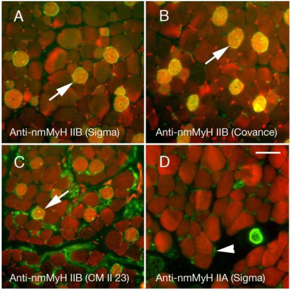
Figure 2.

Adult rat orbits stained for the distribution of nmMyH IIB (green in A-C) using three different antibodies against this protein or IIA (green in D) vs. actin (red in all fields). The polyclonal antibodies are raised against the non-helical tailpiece of myosin and highly label a subset of fibers in the global layers of EOMs (A, B). The monoclonal antibody CM23 II detects nmMyH IIB positive fibers, but labels the neurons better (C). Arrows mark examples of the high level of labeling in A-C. The anti-nmMyH IIA stains satellite cells, neurons, and blood vessels, but nothing within the muscle fibers. Scale bar =10 μm.
