
Figure 2. Phagocytosis of Ig-opsonized sheep-RBC and human-RBC by human-THP-1 macrophages.
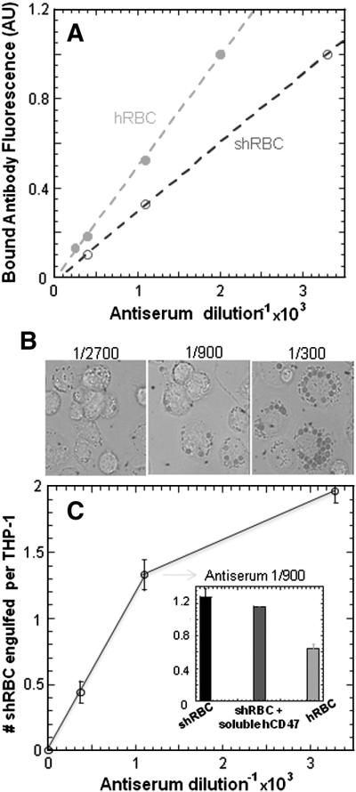
(A) Fresh human and sheep RBC were incubated with antiserum at different dilution ratios and detected by fluorescent secondary antibodies, exhibiting a linear opsonization based on mean fluorescence in flow cytometry. (B) Phagocytosis of shRBC by human-derived THP-1 macrophages at different opsonization level observed by DIC microscopy, which showed a saturable type of response (C). Addition of soluble human-CD47 at μM concentrations showed at most a slight inhibition of phagocytosis of sh-RBC by THP-1 macrophages but not enough to reach hRBC phagocytosis levels at 1/900 antiserum dilution ratio.