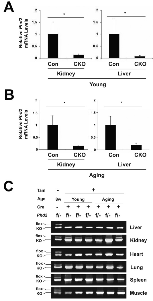
Fig. 2.
Phd2 exon 2 deletion efficiency in mice after tamoxifen induction. Both young and aging mice were treated with tamoxifen for five consecutive days. Four weeks after the initial tamoxifen dose, mice were sacrificed. RNA and DNA were then isolated from select mouse tissues. (A and B) Phd2 mRNA levels in liver and kidney were measured by RT-PCR in (A) young and (B) aging mice. n = 4. * indicates p < 0.05 in comparing control (Con, Phd2 f/+) and CKO (Phd2 f/−; Rosa26-CreERT2) groups at a given age. (C) Phd2 exon 2 recombination efficiency was surveyed in Phd2 CKO mice in both the young and aging groups. A PCR reaction employing three primers was performed on genomic DNA obtained from the indicated tissues. The floxed Phd2 allele without recombination produces a 0.95 kb band, while the knockout Phd2 allele produces a 0.8 kb band. In each panel, the first lane shows results obtained with a control DNA from a Phd2 f/− mouse.

