Fig. 3.
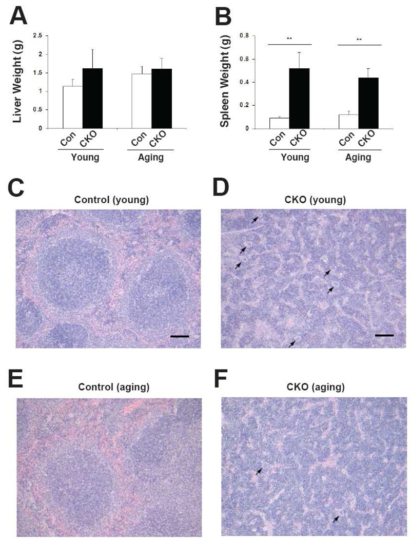
Extramedullary hematopoiesis in Phd2 CKO mice. Both young and aging mice were treated with tamoxifen for five consecutive days. Four weeks after the initial tamoxifen dose, mice were sacrificed. Genotypes are as follows. Controls: Phd2 f/+; CKO: Phd2 f/−; Rosa26-CreERT2. (A) Liver and (B) spleen weights were measured. For (A) and (B), n=6–8. ** indicates p < 0.01. (C–F) Photomicrographs of hematoxylin and eosin stained sections of spleen from young (C,D) and aging (E,F) mice. (Leica DM2500 microscope equipped with Leica FireCam digital capture software, magnification 100×). Bars in (C) and (D) indicate 100 µm. Megakaryocytes are present in the spleens of the Phd2 CKO mice (indicated by arrows). Examination of ten high power fields from two mice in each age group reveals a higher number of megakaryocytes in the spleens of young Phd2 CKO mice (6.3 ± 2.0) as compared to aging Phd2 CKO mice (3.5 ± 1.5; p < 0.01).

