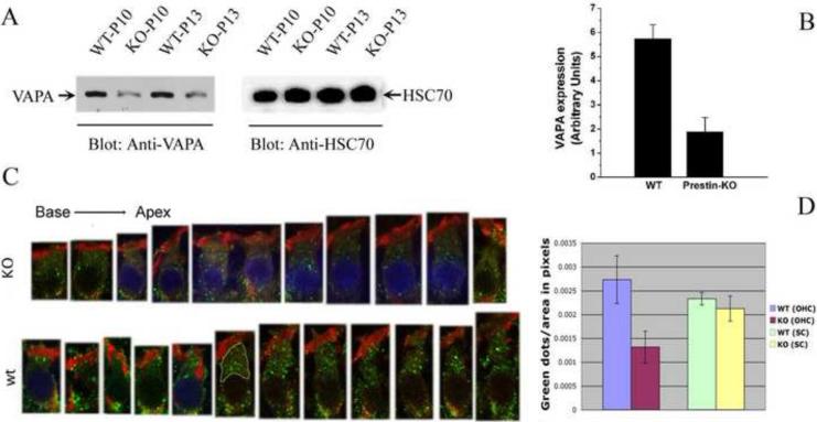
Figure 3.
The expression of VAPA protein is higher in OHCs derived from WT- than from prestin-KO mice. A. Western blot data collected from P10 and P13 cochleae derived from WT- and prestin-KO mice. B. Semi-quantitative analysis of VAPA proteins in WT- and prestin-KO mice. Intensities of VAPA protein bands were divided by band intensities of house-keeping protein HSC70. WT cochlea: n=2, prestin-KO cochlea: n=2 (p=0.01). C. Images of individual OHCs (P13) selected from different locations, either WT-(bottom row) or prestin-KO (top row) mice. D. Quantification of VAPA-staining vesicles (green spots) in WT- and prestin-KO OHCs and supporting cells. The areas between cuticular plates and nuclei, as indicated by a yellow border in one of the WT OHCs in C, were selected and measured using NIH Image J software. More green spots are observed in OHCs from WT-mice, independent of cochlear location. In fact, there is a statistical difference between WT-OHCs and prestin-KO-OHCs (p=0.0001; n=13), with WT-OHCs having more VAPA-green dots per area. The blue staining of some nuclei is due to the mounting of these samples in Vectashield containing DAPI. Other samples, mounted in Fluoromount-G without the DAPI dye, have unstained nuclei. A fixed area in supporting cells of WT- and KO-organ of Corti were selected for counting green spots. In supporting cells, no significant difference (p=0.65) was found in VAPA expression between WT- and KO-cochlea. Green: anti-VAPA. Red: Texas Red-X phalloidin labeling actin.

