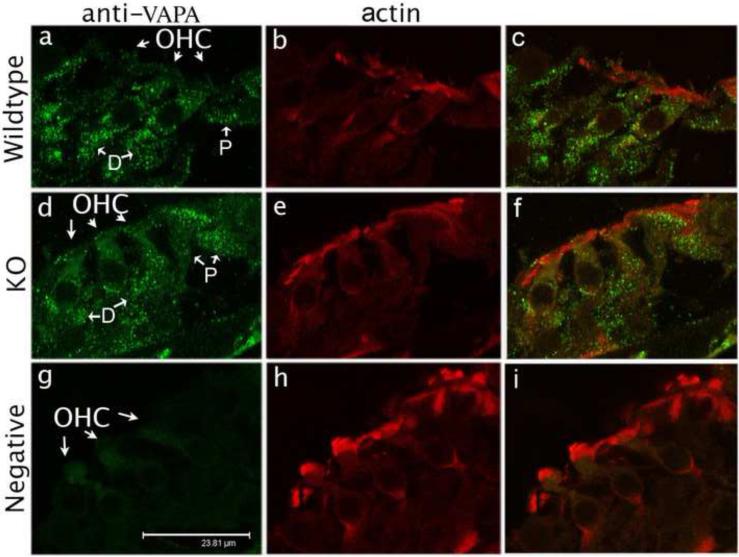
Figure 4.
VAPA expression in the organ of Corti at P13. VAPA staining (anti-VAPA, green spots) is present in OHCs and in supporting cells in both WT (a) and prestin-KO (d) mice. However, punctate VAPA staining is absent in the negative control (g), which is not stained with anti-VAPA primary antibody, but stained with secondary antibody and Texas Red-X phalloidin. Actin (Texas Red-X phalloidin) expression is similar in WT (b), KO (e) and in the negative control (h). The right column (c, f, i) shows superimposed images of first two columns. Bar length: 23.8 μm. D: Deiters’ cells, P: pillar cells.

