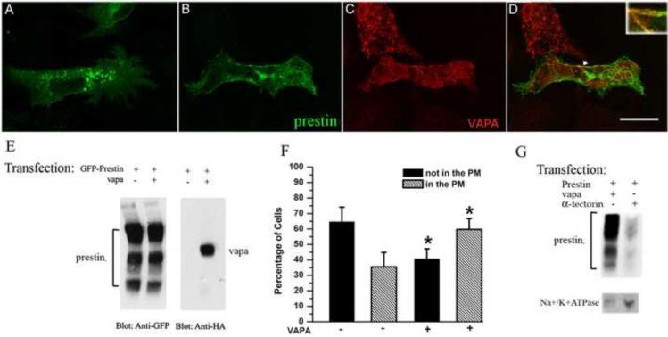
Figure 7.
Prestin distribution patterns under the influence of VAPA. OK cells were transiently transfected with GFP-prestin alone (A) or co-transfected with GFP-prestin+HA-VAPA (B, C). Approximately 28 hrs post transfection, cells were fixed and incubated with anti-HA antibody followed by the corresponding secondary antibody. The yellow image (right column) is superimposed from green and red images, indicating the partial co-localization of prestin and VAPA (D) as indicated by the arrow. For better visualization of the co-localization, the co-localization portion is shown in the right corner at higher magnification. Bar length: 23.8 μm. A shows that prestin is intracellularly localized in the absence of over-expressed VAPA, whereas in the presence of over-expressed VAPA (B) prestin is localized to the plasma membrane. E. Comparison of GFP-prestin expression in OK cells transfected with GFP-prestin alone or co-transfected with GFP-prestin+HA-VAPA. GFP-prestin expression was similar in both GFP-prestin- and GFP-prestin+HA-VAPA transfected cells where GFP-prestin was visualized using anti-GFP antibody. VAPA was observed using anti-HA antibody. F. Distribution of prestin protein under the influence of VAPA. OK cells were transfected with either the plasmid encoding GFP-prestin, or plasmids encoding GFP-prestin+VAPA. OK cells with green staining (prestin) in the PM were defined as “in the PM” (hatched bars). OK cells without green staining in the PM were defined as “not in the PM” (black bars). G. More prestin is delivered to the PM in the presence of over-expressed VAPA than in presence of a control protein, α-tectorin. GFP-prestin was visualized by anti-GFP. Na+/K+ATPase, which is a PM marker, was stained with anti-Na+/K+ATPase.

