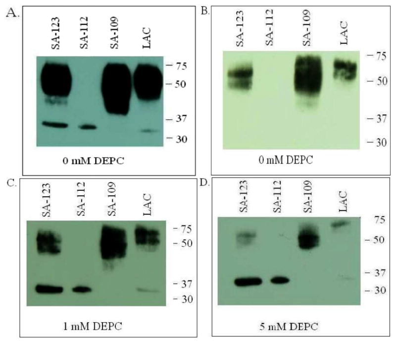
Figure 3. Western blot analysis of culture supernatants from four clinical CA-MRSA isolates.

The isolates vary in the presence of genes for PVL as well as the production of protein A: pvl+ SA-123, pvl+ LAC producing low levels of pvl, pvl- SA-109 and pvl+ SA-112 producing very low levels of Spa. Proteins were separated by SDS/PAGE in 4-20% Tris-glycine gels and transferred to nitrocellulose. Nonspecific sites were blocked by incubating the blots in PBS containing 5% skim milk (PBS-milk). Blots were first incubated with (A) rabbit anti-LukS-PV in PBS-milk, (C) rabbit anti-LukS-PV mixed with 1 mM DEPC, or (D) rabbit anti-LukS-PV mixed with 5 mM DEPC, followed by goat anti-rabbit antibodies conjugated to HRP. (B) protein A was detected using chicken anti-protein A conjugated to HRP. Blots were developed by incubation with Super Signal West Pico Chemiluminescent Substrate and exposure on CL-XPOsure Film.
