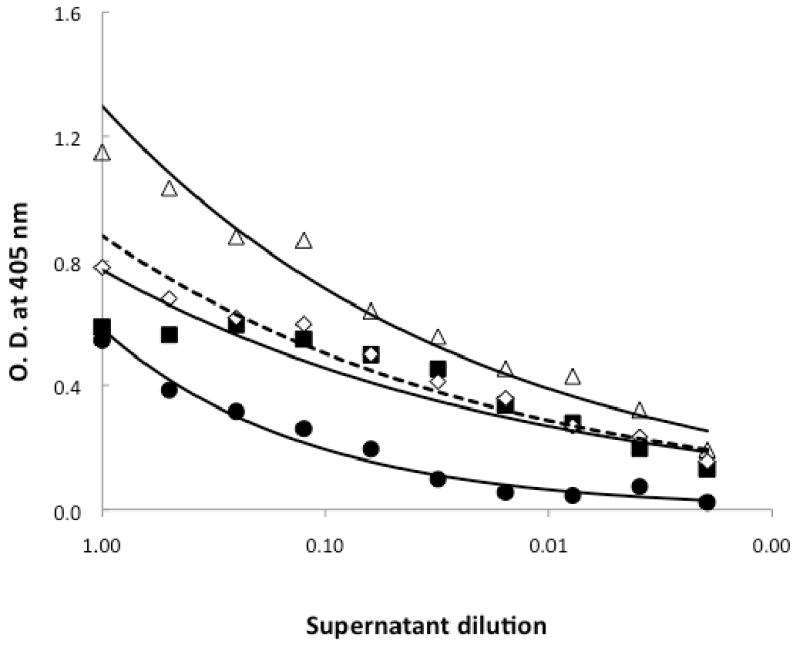
Figure 4. Binding characteristics of anti-LukS-PV pre-incubated with DEPC to rLukS-PV in YCP media and bacterial culture supernatants.

ELISA was carried out as described in Figure 1. Shown is the binding of 2 μg/ml rLukS-PV in dialyzed YCP media to anti-LukS-PV (closed squares, solid lines) and the binding of 2 μg/ml rLukS-PV in dialyzed SA109 supernatant to DEPC-treated anti-LukS-PV (dashed line, open diamonds). Controls shown are dialyzed SA-109 culture supernatants binding to anti-LukS-PV (solid circles) and dialyzed SA-109 culture supernatants containing 2 μg/ml rLukS-PV binding to anti-LukS-PV (open triangles).
