Figure 8.
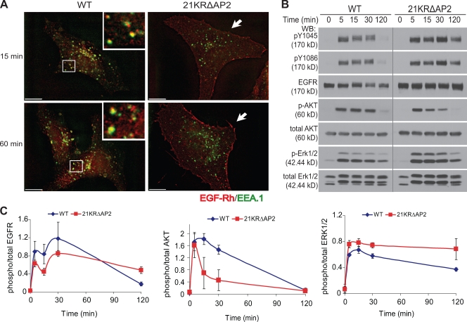
Poor accumulation of 21KRΔAP2 mutant in endosomes and altered kinetics of ERK1/2 and AKT activation by EGF in cells expressing the internalization-defective mutant. (A) PAE cells expressing wtEGFR and 21KRΔAP2 were incubated with 5 ng/ml EGF-Rh for 15 min or 1 h. After fixation, the cells were permeabilized and stained with antibody to EEA.1. A z stack of optical sections was acquired through CY3 (EGF-Rh) and FITC (EEA.1) filter channels and deconvoluted. Insets show enlarged views of the boxed areas, showing localization of wtEGFR in EEA.1-containing early endosomes. 21KRΔAP2 mutant was accumulated in the membrane ruffles (arrows). Bars, 10 µm. (B) Serum-starved cells were treated with 5 ng/ml EGF for 0–120 min at 37°C and lysed. The lysates were probed for active EGFR (pY1045 and pY1086), total EGFR (1005), phosphorylated AKT (p-AKT), total AKT, phosphorylated ERK1/2 (p-ERK1/2), and total ERK1/2. The experiment is representative of three independent experiments. Gray line indicates that intervening lanes have been spliced out. (C) Quantification of experiments presented in B. Bars represent SEM of the amounts of activated EGFR, AKT, and ERK1/2 normalized to total amounts of EGFR, AKT, and ERK1/2, respectively, and plotted against time. The data are averaged from three experiments. WB, Western blot.

