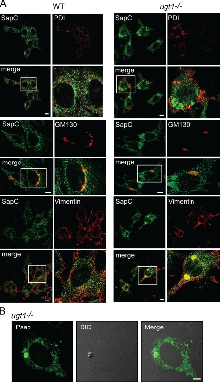
Figure 4.
Prosaposin is localized to aggresome-like inclusions in ugt1−/− cells. Prosaposin localization in wild-type (WT) and ugt1−/− cells was determined by confocal immunofluorescence microscopy. Cells were fixed in 4% paraformaldehyde/PBS and permeabilized in 0.1% Triton X-100. (A) Samples were double labeled with saposin C (SapC) antisera and PDI (ER), GM130 (Golgi), or vimentin (aggresome) antisera. The white boxed regions denote the area magnified in the panels to the right. (B) Samples labeled with full-length prosaposin (Psap) antisera were compared with DIC images. Bars, 10 µm.

