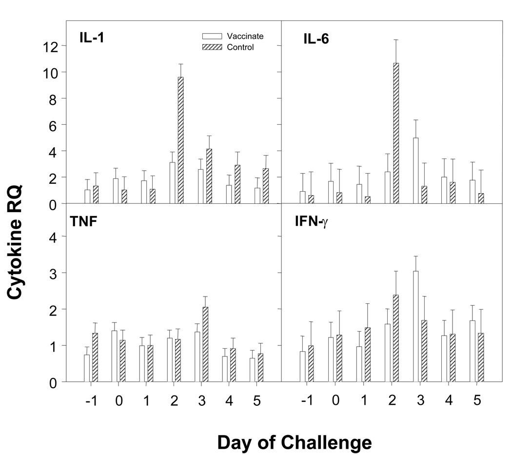
Fig. 5. Pro-inflammatory cytokine mRNA induction post-challenge.
Cytokine mRNA expression in peripheral blood cells was determined by quantitative RT-PCR, using primers/probes as described in Supplementary Materials online(Table 2). Y-axes are relative quantity (RQ) with the average of the Day -1 samples for each cytokine being used as the calibrator. Days post-challenge are shown at bottom. Panels (clockwise from upper left) show mean RQ +/− SEM for IL-1β, IL-6, IFNγ, and TNFα, respectively.

