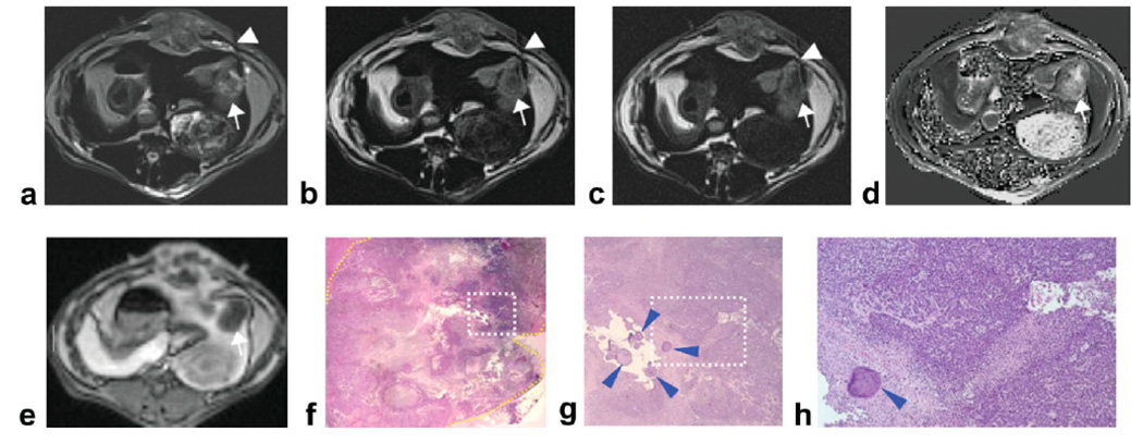
Figure 2.
Left lobe VX2 rabbit liver tumor (diameter = 2.2 cm) (arrows) with intratumoral heterogeneous distribution of viable and necrotic tumor tissues. DW-PROPELLER images (a–c) were acquired with needle (arrowheads) positioned to target the intermediate region at the interface between viable periphery and central necrotic core. Corresponding baseline ADC map shown at right (d). Contrast-enhanced T1W images demonstrate perfusion of the tumor periphery (e). Overview H&E pathology image (×25 magnification) (f) of the tumor (yellow contour line at lesion border) shows central necrotic core with viable tumor periphery and also heterogeneous distribution of mixed tissue types. Magnified image (g) from inset within ×25 overview image (white dashed-box in f) shows the location of the injected beads (blue arrowheads) which served as our ex vivo reference for needle tip position. Tumor tissues within the region of bead deposition (×100 magnification image h from inset position within g) were classified as 50% viable at histopathology.

