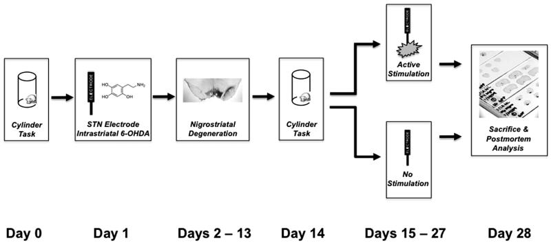
Figure 1. Experimental Design for STN-DBS After Intrastriatal 6-OHDA.
On Day 0 forelimb akinesia was assessed in all rats via the cylinder task. On Day 1 all rats were unilaterally lesioned via injections of 6-OHDA into the striatum and implanted unilaterally (ipsilateral to 6-OHDA) with stimulating electrodes into the STN during the same surgical session. During Days 2-13 all rats were allowed to recover from surgery. On Day 14 all rats were reassessed for degree of contralateral forelimb akinesia via the cylinder task. Rats that displayed a 20% reduction in contralateral forelimb use were divided into two separate groups: ACTIVE (n = 19) or INACTIVE (n = 11) stimulation. Rats in the ACTIVE stimulation group had their stimulators connected to an external stimulation source and received STN stimulation during Days 15-27 twenty-four hours a day. Rats in the INACTIVE group received no stimulation during Days 15-27. On Day 28 all rats were sacrificed and their brains processed for stereological cell counts of THir neurons in the SN, THir neurites in the striatum or levels of DA and DA metabolites in the striatum and frontal cortex. Appropriate placement of the stimulating electrode was confirmed histologically utilizing Kluver-Barrera staining.

