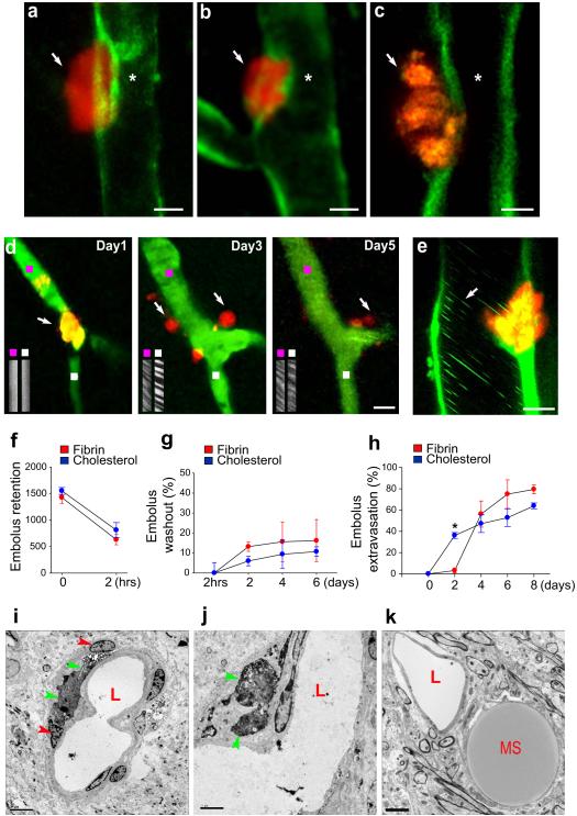
Figure 1. Emboli that fail to be washed-out undergo extravasation leading to blood flow reestablishment.
a-c, Single time point transcranial TPM imaging in Tie2-GFP mice show extravasated fluorescent fibrin clots (a,b arrows; day 4 post-embolization) and a cholesterol embolus (c, arrow; day 3 post-embolization) adjacent to recanalized lumen (asterisk). Scale bars: 10 μm. d, Time-lapse imaging shows a capillary (green; Thioflavin-S dye) occluded by a fibrin clot (orange; arrow, day 1), which extravasates and degrades (arrows, days 3 and 5; Supplementary figure 11). Line-scan imaging upstream (purple squares) and downstream (white squares) of the occlusion shows blood flow reestablishment. e, In vivo image on day 2 shows a cholesterol embolus in the process of extravasation through the GFP-labeled endothelium (ACTB-eGFP mice). Leukocytes (green lines, arrow) are seen flowing even prior to complete extravasation. f, Quantification of fibrin and cholesterol emboli (10-20 μm) retained in the microvasculature which failed to be lysed or washed-out 2 hours post-embolization (~1500 clots per mouse in 12 mice). g, Fibrin and cholesterol emboli washout up to 6 days postembolization (mean ± s.e.m. n=3 mice per time point). h, Fibrin and cholesterol emboli extravasation up to 8 days post-embolization (mean ± s.e.m.; n=10 mice and 17 fibrin clots and n= 10 mice and 18 cholesterol emboli per mouse). The difference in early extravasation rates between cholesterol and clots (asterisk, p<0001) is likely due to a tendency of clots to dislodge from their initial site of occlusion. i-k, Transmission electron microscopy (TEM) shows (i,j), colloidal carbon-conjugated fibrin clots (green arrowheads) which have extravasated after 7 days and are surrounded by the processes of perivascular cells (red arrowheads) and (k), a microsphere (MS) outside the capillary lumen (L). Scale bars 5μm.

