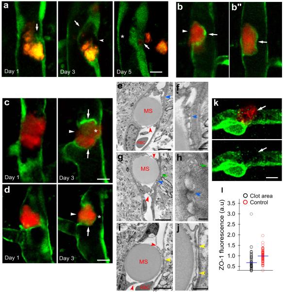
Figure 2. Focal endothelial remodeling underlies the translocation of emboli.
a, Time lapse imaging in Tie2-GFP mice shows gradual extension of a membrane from the adjacent endothelium (arrow, day1) eventually surrounding a cholesterol embolus (orange) (arrow, day 3). The original endothelium undergoes retraction (arrowhead, day 3) creating a path for embolus translocation. On day 5, the embolus has extravasated leading to lumen recanalization (asterisk). b,b”, Single time point in vivo image shows two optical planes of the same vessel. The fibrin clot (red) is enveloped by a newly formed endothelial extension (arrow) while an opening in the endothelium is visible through which the clot appears to be extravasating (b, arrowhead) (Supplementary Movie 1). c,d, Time lapse images in Tie2-GFP mice show fibrin clots (red) at different stages in the formation of the endothelial envelope (arrows), endothelial retraction (arrowheads) and lumen reestablishment (asterisks). e-j, TEM shows intravascular microspheres which have been enveloped by endothelial extensions (red arrowheads). The newly formed membranes make contact with the opposing endothelium forming specialized cellular junctions (e-h; blue arrowheads) and small vacuolar structures (g,h; green arrows). (i-j), Endothelial membranes are detached from each other and a narrow lumen is apparent (yellow arrowheads). Red blood cells (RBC). k, Confocal image of a microvessel immunolabeled for Zonula Occludens-1 (ZO-1) (green) shows reduced labeling (arrows) adjacent to intravascular fibrin clot (red). Scale bar 10μm. l, Quantification of ZO-1 immunofluorescence adjacent to clots and control unoccluded microvessels in the same confocal optical plane (n=5 mice and 55 and 57 vessels, p<0.001, two-side Mann-Whitney test).

