
Figure 6.
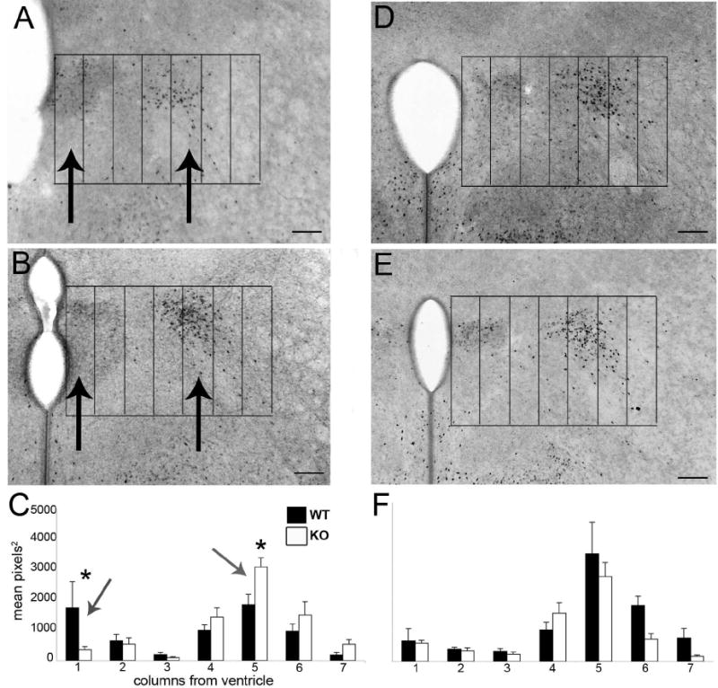
Digital mages show ERα immunoreactivity at P0 in GABABR1 subunit wildtype (A,D) and knockout (B,E) mice in the plane of section corresponding to figure 4B. Seven 100μm wide columns were placed over images with the edge of the third ventricle and the top of the paraventricular nucleus as the boundaries for the grid. Female GABABR1 subunit knockouts (n=6) had an approximately 80% decrease in immunoreactivity in column 1, as measured by mean pixels2, the first 100μm from the ventricle (Nguyen-Ba-Charvet et al., 2004), and an approximately 70% increase in immunoreactive area in column 5 (400-500μm from the ventricle), versus wildtype littermates (n=6). There was no difference in levels of immunoreactivity in male GABABR1 subunit knockout mice (D-F; n=6) as compared to wildtype (n=6) (scale bars =100μm).