
Figure 2. Loss of CD95 expression inhibits ovarian cancer in vivo.
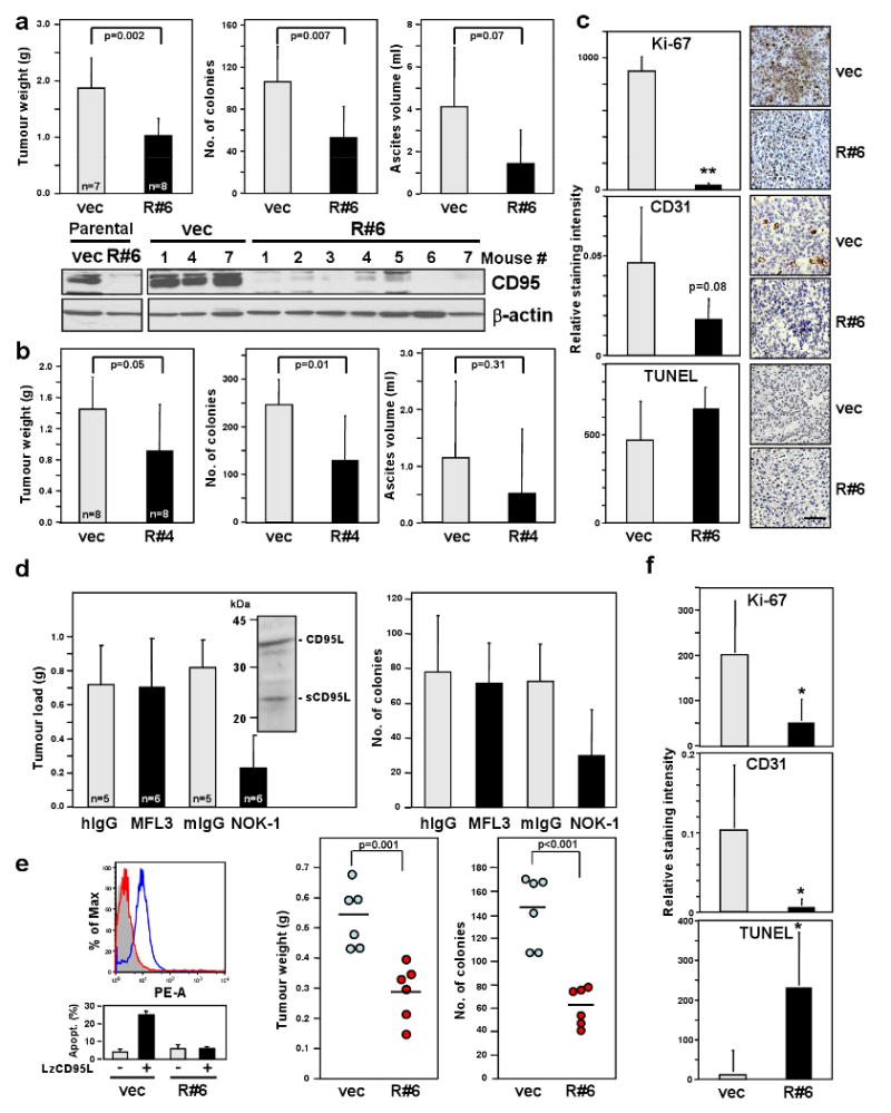
a Tumour weight, number of tumour colonies and ascites from mice injected with SKOV3ip1 vec or R#6 cells. Lysates of cells and tumour tissues were examined for CD95 level by western blot analysis. b, Same parameters as in (a) were measured from mice injected with SKOV3ip1 vec or R#4 cells. c, Histology and immunohistochemistry staining for Ki-67, TUNEL and CD31 of SKOV3ip1 vec and R#6 tumours. Scale bar = 100 μm. ** p-value <0.001. d, Tumour load and number of tumour colonies of mice treated with neutralising mAb for murine CD95L (MFL3), human CD95L (NOK-1) or corresponding isotype control mAbs were measured. Inset, Western blot analysis of SKOV3ip1 cell lysate for CD95L. e, Surface CD95 staining (upper left) and apoptosis sensitivity by LzCD95L (100 ng/ml) treatment (lower left) of MONTY-1 vec and R#6 cells. Weight and number of colonies of tumours formed by MONTY-1 vec and R#6 cells are shown (right). f, The staining intensity for Ki-67, TUNEL and CD31 of tumours from MONTY-1 vec and R#6 cells were quantified. * p-value <0.05. Values in graphs in a to e and f represent mean -/+ s.d. from three independent experiments. The horizontal bars in right part part of e represent the mean of 6 animals.