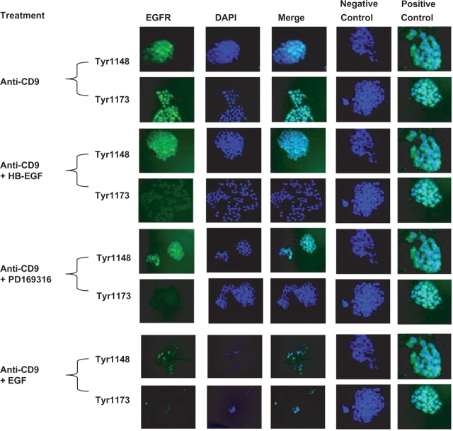
Fig. 3.
Phosphorylation of EGFR is suppressed by HB-EGF and p38 MAPK inhibitor (20×). After 24 h of anti-CD9 antibody treatment and fixation cells were stained for activated EGFR 1148 and 1173 (green). Nuclei are labelled with DAPI. Anti-CD9 antibody treatment induced phosphorylation of EGFR Y1148 and Y1173. HB-EGF and p38 inhibitor suppressed the activation of Y1173 while EGF did not as significantly. Matching isotype control antibody did not activate any of the EGFR residues of interest (negative control). Separately treated cells with EGF or HB-EGF produced activated EGFR (positive control).

