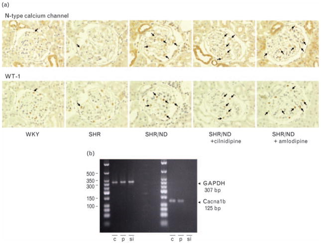
Fig. 2.
The co-expression of N-type calcium channel with podocyte-specific nuclear protein, WT-1 (a) and the expression of N-type calcium channel in cultured podocyte (b). The representative glomerulus pictures from WKY and SHR/ND were shown. The immunoreactivities for both N-type calcium channel and WT-1 were found in the same cells (arrows) analyzed in the consecutive kidney sections (1.5 μm thick). Reverse-transcription PCR analysis revealed significant expression of the N-type calcium channel product (125 bp) in podocyte (p) and cerebral cortex (c) as a positive control, and the band was abolished by siRNA transfection of N-type calcium channel (si) with the same proportion of GAPDH expression (307 bp) (n = 3). SHR, spontaneously hypertensive rat; WKY, Wistar–Kyoto rats; WT-1, Wilms’ tumor factor 1.

