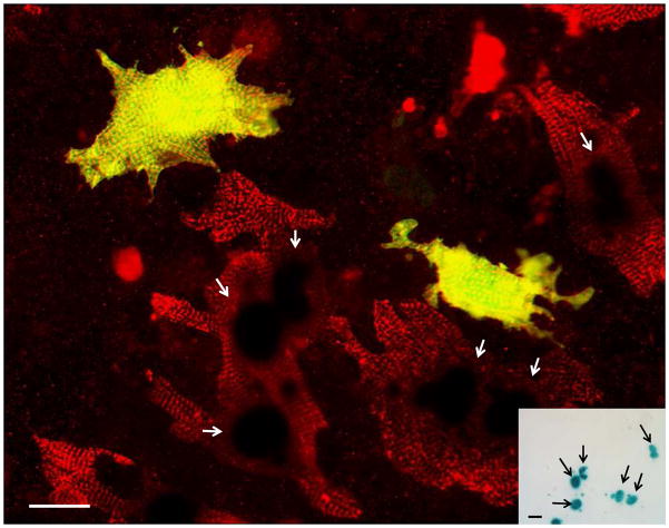
Figure 4. Cardiac-resident c-kit+ cells from neonatal hearts exhibit robust cardiomyogenic differentiation in the absence of cell fusion.
Fetal cardiomyocyte cultures from single transgenic MHC-nLAC mice were seeded with c-kit+ cells from neonatal ACT-EGFP hearts and cultured for 7 days. The cultures were then fixed and processed for β-galactosidase activity (blue), EGFP (green) and α-actinin (red) immune reactivity. EGFP-expressing cardiomyocytes exhibiting mature sarcomeric structure (yellow due to the overlay of green EGFP and red α-actinin signals) were readily detected by fluorescence microscopy; however these cells lacked β-galactosidase activity when visualized under bright field illumination (inset). Bar = 20 microns in the fluorescent image, 20 microns in the bright field inset. Arrows mark β-galatosidase positive nuclei. Bright field, single color fluorescence and merged images for these cells are shown in the Supplemental Data section.

