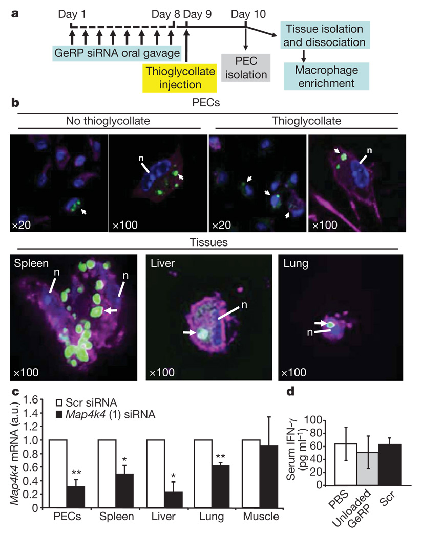
Figure 4. Orally administered GeRPs containing Map4k4 siRNA attenuate Map4k4 mRNA expression in PECs and macrophages from spleen, lung and liver.
a, Time line of GeRP (scrambled (Scr) or Map4k4, oligo 1) administration and PEC/tissue isolation. b, Confocal microscopy of PECs and tissue macrophages containing GeRPs (green). Macrophages were stained with F4/80–AF633 (magenta). Arrows indicate cells containing GeRPs. Nuclei (n) were stained with 4,6-diamidino-2-phenylindole (DAPI, blue). Magnification is indicated in the panels. c, Map4k4 mRNA expression in PECs and adherent cells from tissues. d, Serum INF-γ levels from mice gavaged with PBS, unloaded GeRPs or GeRPs containing 20 µg kg−1 of Scr siRNA (n = 5). Statistical significance was determined by ANOVA and Tukey post test. **P < 0.01 and *P < 0.05. Results are mean ± s.e.m.

