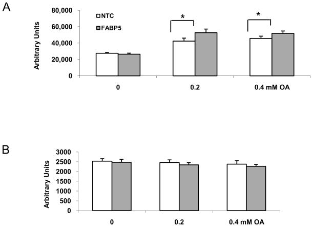
Figure 4.
The levels of neutral lipids measured by Nile Red staining. A. After treated with siRNA for 6 h, the cells were incubated in the medium containing 1% BSA for 24 h. Nile Red assay was performed after cells incubated in medium supplemented with 0–0.4mM oleic acid for an additional 24 h. B. Cytotoxicity assays. The cells were incubated with 10 μM of resazurin in HBSS. The background fluorescence was read twice, with the fluorescence being recorded at 560/590 nm: immediately and after 1 h of incubation at room temperature. Data shown are means ± SD from four replicates of one representative experiment from three independent experiments. *p < 0.05 relative to controls.

