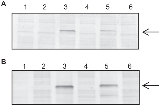
Figure 4.
Detection of cPKA Fusion Proteins Expressed in RR Before and After Immobilization. A) Western Blot Analysis. The amount of fusion protein before and after immobilization was determined by gel electrophoresis followed by immunoblotting with antibody specific for PKA at a 1:1000 fold dilution. A volume equivalent to 1 μl of the protein synthesis reaction was loaded per lane. Lanes 1 and 2: no DNA template negative control reaction; Lanes 3 and 4: pFN19-cPKA; Lanes 5 and 6: pFC20-cPKA. The starting materials from the TNT reactions are in lanes 1, 3 and 5, and the post-immobilization supernatants (“flowthroughs”) are in lanes 2, 4 and 6. B) HaloTag TMR Ligand Binding Assay. The starting material and post-immobilization supernatants were incubated with HaloTag TMR Ligand as indicated in the Experimental Section. A volume equivalent to 1 μl of the starting reaction was loaded per lane in the same lane order listed in Panel A. A comparison of protein levels in lanes 3 and 4, and lanes 5 and 6, is made to determine binding of the fusion protein to the magnetic beads. The arrow points to the expressed cPKA fusion protein.

